Intra Oral Image of Dental Fluorosis
Amal H Abuaffan
Amal H Abuaffan*
Department of Orthodontic, Pedodontics and Preventive Dentistry, Faculty of Dentistry, Khartoum University, Khartoum, Sudan
- *Corresponding Author:
- Amal H Abuaffan
Department of Orthodontic, Pedodontics and Preventive Dentistry
Faculty of Dentistry, Khartoum University
Khartoum, Sudan
Tel: +249912696035
E-mail: amalabuaffan@yahoo.com
Clinical Presentation
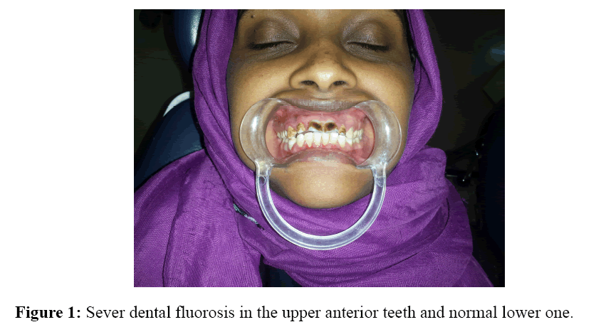
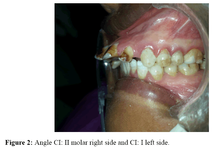
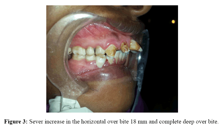
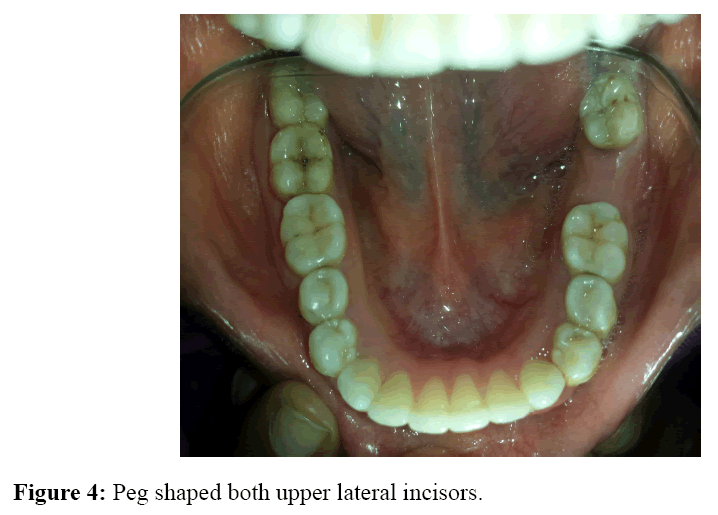
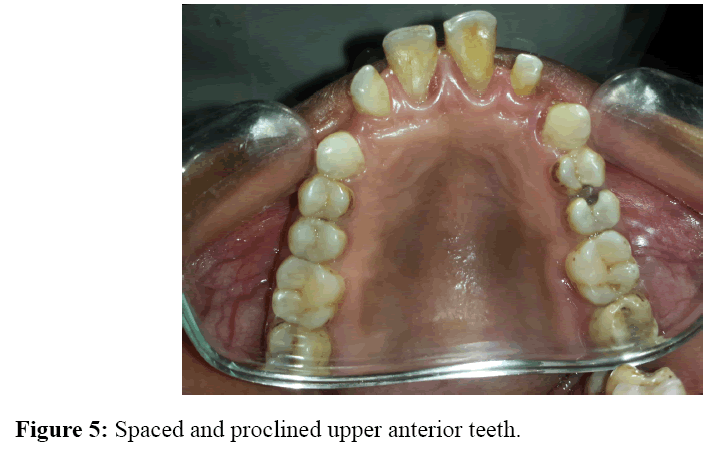
Intra oral image for Sudanese lady 25 years old with the following problems: Intra oral image for Sudanese lady 25 years old with the following problems: Sever dental fluorosis in the upper anterior teeth and normal lower one. Angle CI: II molar right side and CI: I left side. Sever increase in the horizontal over bite 18 mm and complete deep over bite. Peg shaped both upper lateral incisors (Figures 1-5).
Figure 1: Sever dental fluorosis in the upper anterior teeth and normal lower one.
Figure 2: Angle CI: II molar right side and CI: I left side.
Figure 4: Peg shaped both upper lateral incisors. Figure 5:
Figure 5: Spaced and proclined upper anterior teeth.
Open Access Journals
- Aquaculture & Veterinary Science
- Chemistry & Chemical Sciences
- Clinical Sciences
- Engineering
- General Science
- Genetics & Molecular Biology
- Health Care & Nursing
- Immunology & Microbiology
- Materials Science
- Mathematics & Physics
- Medical Sciences
- Neurology & Psychiatry
- Oncology & Cancer Science
- Pharmaceutical Sciences
