Spectrum of Micturating Cystourethrogram Revisited: A Pictorial Assay
Abhinav Jain, Vivek Setia, Shweta Agnihotri
Abhinav Jain1*, Vivek Setia1, Shweta Agnihotri2
1Department of Radio Diagnosis, Hamdard Institute of Medical Sciences and Research, Jamia Hamdard, New Delhi, India
2Bansal Hospital, New Friends Colony, New Delhi, India
- *Corresponding Author:
- Abhinav Jain
Department of Radio Diagnosis
Hamdard Institute of Medical Sciences and Research
Jamia Hamdard, New Delhi, India
Tel: +919999912701
E-mail: drabhinavjain@gmail.com
Abstract
Micturating Cystourethrography (MCU) is a useful technique to evaluate the abnormalities of urinary tract. With advent of newer imaging techniques, conventional radiography is largely being abandoned; however cystourethrography is still a preferred imaging technique for assessment of urinary bladder, urethra and VUR especially in children. We present a spectrum of abnormalities on MCU studies affecting urethra, urinary bladder and ureter seen over a period of three years in radiology department of our teaching hospital.
Keywords
Cystourethrogram, Cystourethrography
Introduction
Micturating cystourethrography (MCU), also known as Voiding cystourethrography (VCUG), is a fluoroscopic study of the lower urinary tract mainly to assess the urinary bladder, urethra, postoperative anatomy and micturition to assess bladder and urethral abnormalities, including vesicoureteric reflux (VUR) with very low radiation exposure if done properly.[1] With radiological advancements the newer techniques like voiding urosonography,[2] nonionising photoacoustic cystography [3] and CT cystography for bladder trauma [4] may soon supersede MCU. However, at present MCU remains mainstay in assessment of these structures, particularly in children.[4] MCU is commonly indicated for following abnormalities [5]:
Vesicoureteric reflux (VUR) including follow up evaluation of VUR.[6]
Study of the urethra during micturition for strictures, posterior urethral valves or urethral trauma.s
Abnormalities of the bladder like diverticulum, foreign bodies and fistulas, bladder outlet obstruction, post traumatic bladder rupture, neurologic bladder.
Stress incontinence.
Congenital anomalies of genitourinary tract.
It is also important to stress that acute urinary tract infection is a contraindication for this study.[7] Most of the recommendations are for an interval of at least 3 to 6 weeks after a UTI before performing MCU.
Methods
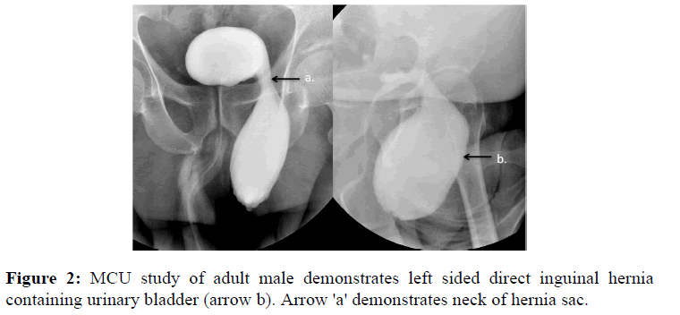
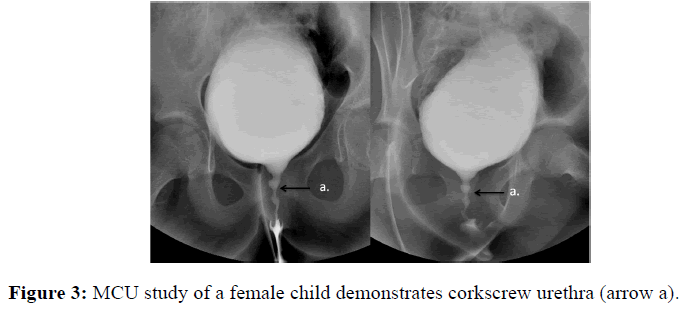
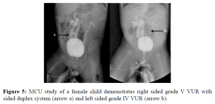
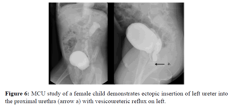
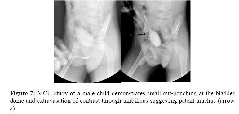
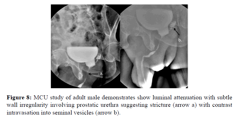
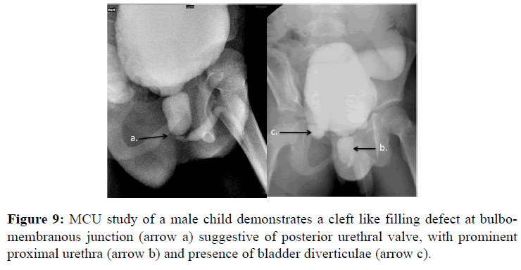
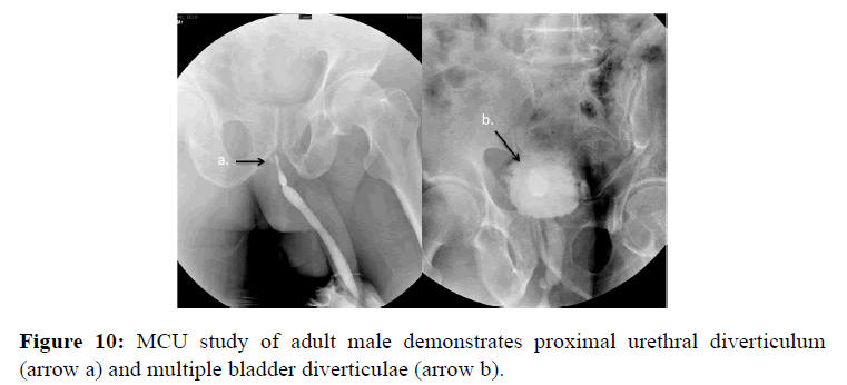
More than 100 MCUs were done in last three years at our hospital. These studies were retrospectively retrieved and reviewed by 3 experienced radiologists. The images depicting key abnormalities were selected and annotated to prepare this pictorial assay. Literature was systematically reviewed using standard search engines to understand the key radiographic abnormalities on MCU studies, various indications and methods of MCU study (Figures 1-10).
Conclusion
Knowledge of imaging spectrum for MCU studies helps in identifying the uncommon abnormal appearances and hence proper management of the pathologies.
References
- Leibovic SJ., Lebowitz RL. Reducing patient dose in voiding cystourethrography. Urol Radiol 1980; 2: 103-107.
- Darge K. Voiding urosonography with US contrast agent for the diagnosis of vesicoureteric reflux in children: an update. Pediatr Radiol 2010; 40: 956-962.
- Kim C., Jeon M., Wang LV. Nonionizing photoacoustic cystography in vivo. Opt Lett 2011;36:3599-3601.
- Ishak C., Kanth N. Bladder trauma: multidetector computed tomography cystography. Emerg Radiol 2011; 18:321-327.
- Jequier S., Jequier JC. Reliability of voiding cystourethrography to detect reflux. AJR Am J Roentgenol 1989; 153: 807-810.
- Lebowitz RL.The detection and characterization of vesicoureteral reflux in the child.J Urol1992; 148:1640-1642.
- Shaikh N., Craig JC., Rovers MM. Identification of children and adolescents at risk for renal scarring after a first urinary tract infection: a meta-analysis with individual patient data. JAMA Pediatr 2014; 168: 893-900.
Open Access Journals
- Aquaculture & Veterinary Science
- Chemistry & Chemical Sciences
- Clinical Sciences
- Engineering
- General Science
- Genetics & Molecular Biology
- Health Care & Nursing
- Immunology & Microbiology
- Materials Science
- Mathematics & Physics
- Medical Sciences
- Neurology & Psychiatry
- Oncology & Cancer Science
- Pharmaceutical Sciences

