Duplication of Gall Bladder A Rare Anomaly
Rajul Rastogi, Yuktika Gupta, Pragya Sinha, Pankaj Kumar Das, Mohini Chaudhary, Vijai Pratap
Rajul Rastogi*, Yuktika Gupta, Pragya Sinha, Pankaj Kumar Das, Mohini Chaudhary and Vijai Pratap
Teerthanker Mahaveer Medical College & Research Centre, Moradabad, Uttar Pradesh, India
- *Corresponding Author:
- Rajul Rastogi
Teerthanker Mahaveer Medical College & Research Centre
Moradabad, Uttar Pradesh, India
E-mail: eesharastogi@gmail.com
Abstract
Duplication of gall bladder (GB) or double GB is a rare congenital anomaly often missed on imaging and during operative procedure. Recognition of this anomaly has gained more significance in the present era of laparoscopic and robotic surgeries as the diseases may involve one or both of them affecting prognosis. In this article, we present a case of duplicated gall bladder that was diagnosed confidently on ultrasonography.
https://sporbahisleri.blogaaja.fi http://sporbahisleri.parsiblog.com https://spor-bahisleri.jimdosite.com https://sporbahisleri.edublogs.org https://sporbahisleri.websites.co.in https://sporbahisleri.podia.com https://sporbahisleri7.wordpress.com https://sporbahisleri.jigsy.com https://niwn-chroiaty-mcieung.yolasite.com https://spor-bahisleri.mywebselfsite.net https://sporbahisleri.mystrikingly.com https://sporbahisleri.splashthat.com https://sporbahisleri1.webnode.com.tr https://sporbahisleri.odoo.com http://sporbahisleri.creatorlink.net http://www.geocities.ws/sporbahisleri/ https://spor-s-site.thinkific.com https://artistecard.com/sporbahisleri https://sporbahisleri.estranky.cz https://spor-bahisleri.mozellosite.com https://651be6b563e56.site123.me https://betsitesiinceleme.blogspot.com https://sporbahisleri.hashnode.dev https://sporbahislerim.wixsite.com/spor-bahisleri https://sporbahislerix.weebly.com https://sites.google.com/view/betsiteleri https://codepen.io/sporbahisleri https://sporbahisleri.bcz.com https://www.smore.com/6rsb9
Keywords
Duplication, Double, Gall bladder, Ultrasonography
Case Report
A 5-year old male child with a history of pain abdomen and mild jaundice came for ultrasonography (USG) of abdomen. His SGPT was raised measuring up to 950 IU/ml associated with raised total bilirubin (3.5% mg) with predominantly raised direct bilirubin (2.3% mg).
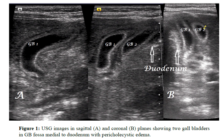
USG abdomen revealed two similar-appearing, gall-bladder like structures in the gall bladder fossa lying adjacent to each other near the duodenum with diffuse mural thickening (measuring maximally up to 5 mm) and pericholecystic fluid without obvious intraluminal focal lesion (Figures 1A and 1B). No evidence of any other sonographic abnormality was noted. Based on the above findings, true duplication of gall bladder (GB) with signs of acute hepatitis was made.
As patient did not have any symptomatic disease, hence patient was just advised follow-up.
Discussion
Duplication of GB is an unusual & rare anomaly of biliary tree with an estimated incidence of one in 4000 in post-mortem studies. [1] It is often missed on imaging studies as well as surgeries due to lack of awareness about this entity especially when one of them is diseased and other normal. [2]
Duplication of GB is of two types according to Boyden’s classification: [3]
Bilobed or vesica fellea divisa
True duplication or vesical fellea duplex with two cystic ducts.
True duplication is further subdivided in to V-type where two cystic ducts unite before opening in to the common bile duct and H-type or ductular-type where they open separately in to common bile duct.
In true duplication, the two GB are usually noted in their physiological infrahepatic location adjacent to each other sharing same peritoneal covering except in few cases where one of them may be intrahepatic or subhepatic.1 Similarly, cystic arteries may be single or duplicated.
There are no specific symptoms of duplicated GB. Diseases like cholelithiasis, cholecystitis & carcinoma may affect one or both GB simultaneously or one after another making preoperative recognition of this rare entity imperative. Failure of recognition on imaging or surgery carries a high risk of injury to biliary tree with recurrent cholecystitis as has been reported in some previous studies. [1,4]
The advent of modern imaging has made confident preoperative recognition of this rare entity possible as seen in our case. However, though ultrasonography (USG), computed tomography (CT) and magnetic resonance imaging (MRI) may allow a confident diagnosis yet they are not 100% sensitive as this rare anomaly requires differentiation from commoner conditions including choledochal cyst, GB diverticulum, and fibrous bands across GB and rarely folded GB.5 Intraoperative cholecystogram may be useful in delineating complete biliary anatomy in suspected cases. [1]
Recognition of this rare entity at imaging or surgery is important not only because removal of accessory or second GB is recommended to avoid postoperative complications & morbidity related to re-explorations but also to prevent unnecessary intervention in asymptomatic individuals where this anomaly may be confused with other variations/pathologies. [1]
Very few cases of preoperative diagnosis on imaging have been reported in literature with few of them showing duplicated GB on antenatal USG as well. [6,7]
Conclusion
Duplication of GB is a rare biliary tract anomaly that should be recognised by the imaging experts to serve as a roadmap for operating surgeon in order to prevent undesirable operative and postoperative morbidity.
References
- Vijayaraghavan R., Belagavi CS. Double gallbladder with different disease entities: a case report. J Minim Access Surg 2006; 2: 23-26.
- Mazziotti S., Minutoli F., Blandino A., Vinci S., Salamone I., et al. Gallbladder duplication: MR cholangiography demonstration. Abdom Imaging 2001; 26: 287-289.
- Boyden EA. The accessory gallbladder, an embryological and comparative study of aberrant biliary vesicles occurring in man and domestic mammals. Am J Anesthesiol 1926; 38: 177-231.
- Yasir M., Kapoor M., Ajman A., Suri A. Double gall bladder: a rare anomaly diagnosed during the laparoscopic cholecystectomy. J SciSoc 2014; 41: 45-46.
- Horattas MC. Gallbladder duplication and laparoscopic management. J LaparoendoscAdvSurg Tech 1998; 8: 231-235.
- Desolneux G., Mucci S., Lebigot J., Arnaud JP., Hamy A. Duplication of the Gallbladder–a Case report. Gastroenterology Research and Practice 2009; 4: 3.
- Kinoshita LL., Callen PW., Filly RA., Hill LM. Sonographic detection of Gallbladder Duplication. J Ultrasound Med 2002; 21: 1417–1421.
Open Access Journals
- Aquaculture & Veterinary Science
- Chemistry & Chemical Sciences
- Clinical Sciences
- Engineering
- General Science
- Genetics & Molecular Biology
- Health Care & Nursing
- Immunology & Microbiology
- Materials Science
- Mathematics & Physics
- Medical Sciences
- Neurology & Psychiatry
- Oncology & Cancer Science
- Pharmaceutical Sciences

