Left Atrial Myxoma: Echocardiographic and CT Images
Ranjit Meher, Donboklang Lynser, Taraprasad Tripathy
Ranjit Meher, Donboklang Lynser*, Taraprasad Tripathy
Radiodiagnosis and Imaging, North Eastern Indira Gandhi Regional Institute of Health and Medical Sciences, Shillong 793018, Meghalaya, India
- *Corresponding Author:
- Donboklang Lynser
Radiodiagnosis and Imaging
North Eastern Indira Gandhi Regional Institute of Health and Medical Sciences
Shillong 793018, Meghalaya, India
Tel: 00919774655769
E-mail: bokdlynser@yahoo.co.in
Clinical Presentation
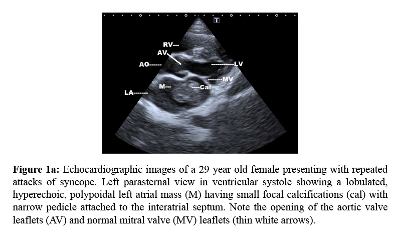
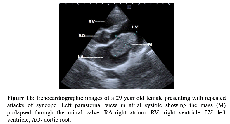
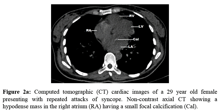
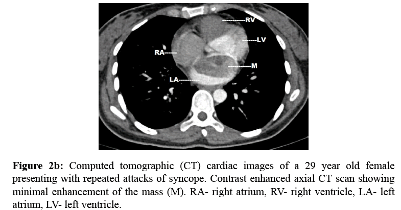
A 29-year-old female was evaluated for repeated attacks of syncope. Echocardiographically, a large, well defined, polypoidal, lobulated hyperechoic mass measuring 4.7 cm × 2.5 cm having a focal area of calcification in a dilated left atrium (Figure 1a). The lesion has a narrow pedicle to interatrial septum near fossa ovalis and seen prolapsing into the left ventricle during atrial systole (Figure 1b). Plain CT thorax revealed a hypodense soft tissue mass with a focal calcification (Figure 2a). On CECT there is minimal enhancement ruling out thrombus (Figure 2b). These features are consistent with left atrial myxoma.
Figure 1a: Echocardiographic images of a 29 year old female presenting with repeated attacks of syncope. Left parasternal view in ventricular systole showing a lobulated, hyperechoic, polypoidal left atrial mass (M) having small focal calcifications (cal) with narrow pedicle attached to the interatrial septum. Note the opening of the aortic valve leaflets (AV) and normal mitral valve (MV) leaflets (thin white arrows).
Primary cardiac tumours in adults are rare. Myxoma is the most common cardiac tumour, accounting for 50% of all cases. 75% of cardiac myxomas occur in the left atrium, 20% in right atrium and rest are in ventricles. It can present with embolic phenomena, congestive cardiac failure, chest pain, pulmonary hypertension, syncope, arrhythmia and systemic symptoms such as fever, malaise and weight loss [1].
Echocardiographically cardiac myxomas usually present as lobulated, hyperechoic, polypoidal left atrial mass with narrow pedicle to interatrial septum near fossa ovalis [2]. 14% show coarse or punctate calcification. On CT scan, 95% myxomas are spherical or ovoid, 76% lobular contour and 24% smooth. On CECT chest, 81% myxomas are hypodense, 19% isodense and 67% heterogeneous [3]. Cardiac tumour including atrial myxoma must be ruled in patient with repeated attacks of syncope. Echocardiography is the most useful primary imaging tool and CECT is the most efficacious imaging to differentiate myxoma from thrombus.
References
- Silverman NA. Primary Cardiac Tumors. Annals of surgery 1980; 191: 127-138.
- Blanchard DG., DeMaria AN. Cardiac and extra-cardiac masses: echocardiographic evaluation. In: Skorton DJ, eds. Marcus cardiac imaging: a companion to Braunwald’s heart disease. 2nd ed. Philadelphia, PA: Saunders 1996; 452-480.
- GrebencML., Rosado-de-Christenson ML., Green CE., Burke AP., Galvin JR. Cardiac Myxoma: Imaging Features in 83 Patients. RadioGraphics 2002; 22: 673–689.
Open Access Journals
- Aquaculture & Veterinary Science
- Chemistry & Chemical Sciences
- Clinical Sciences
- Engineering
- General Science
- Genetics & Molecular Biology
- Health Care & Nursing
- Immunology & Microbiology
- Materials Science
- Mathematics & Physics
- Medical Sciences
- Neurology & Psychiatry
- Oncology & Cancer Science
- Pharmaceutical Sciences


